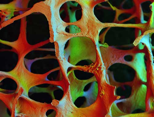
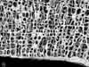
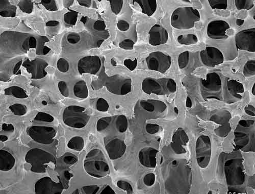
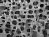
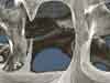
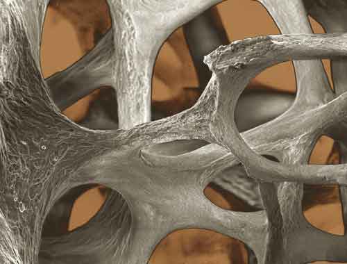
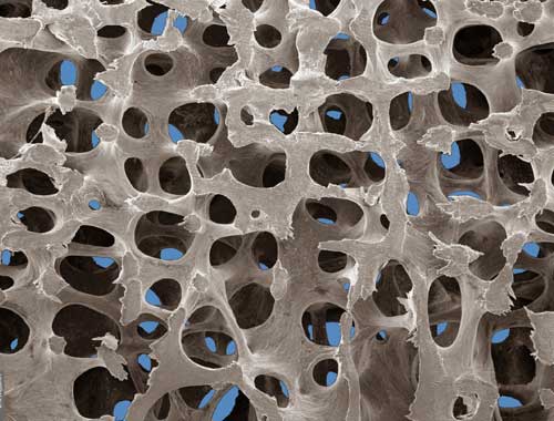
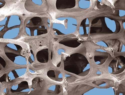
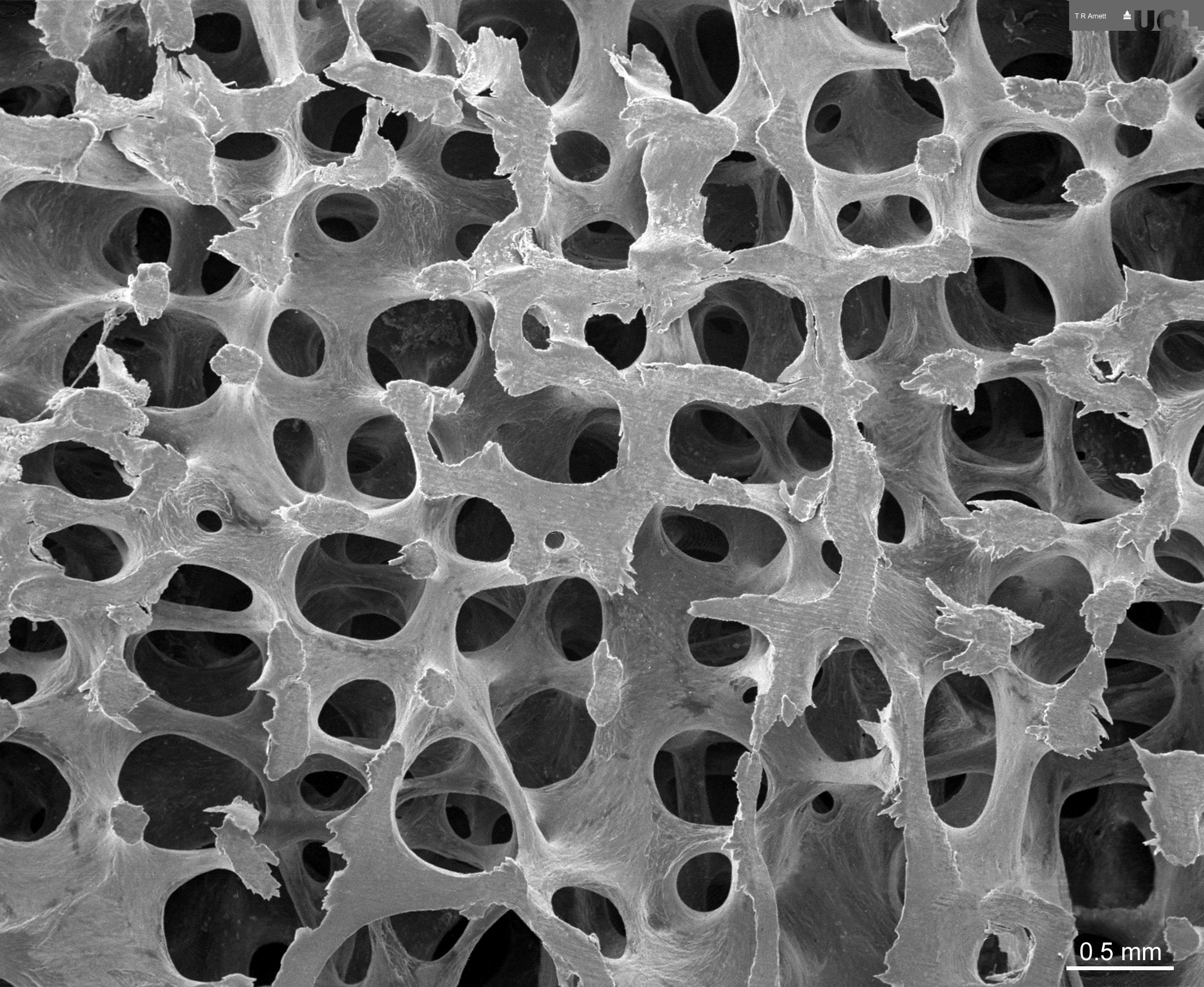
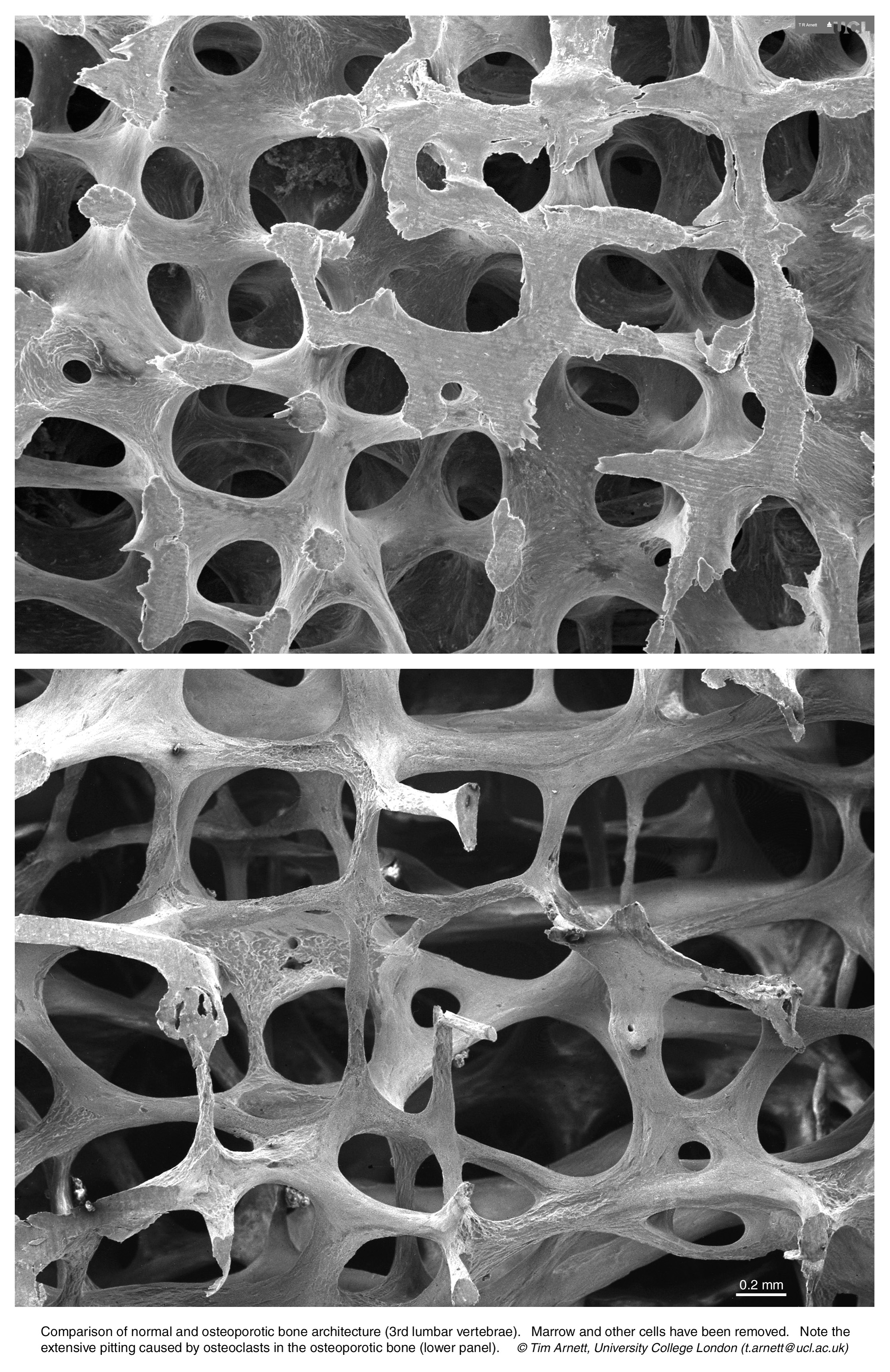
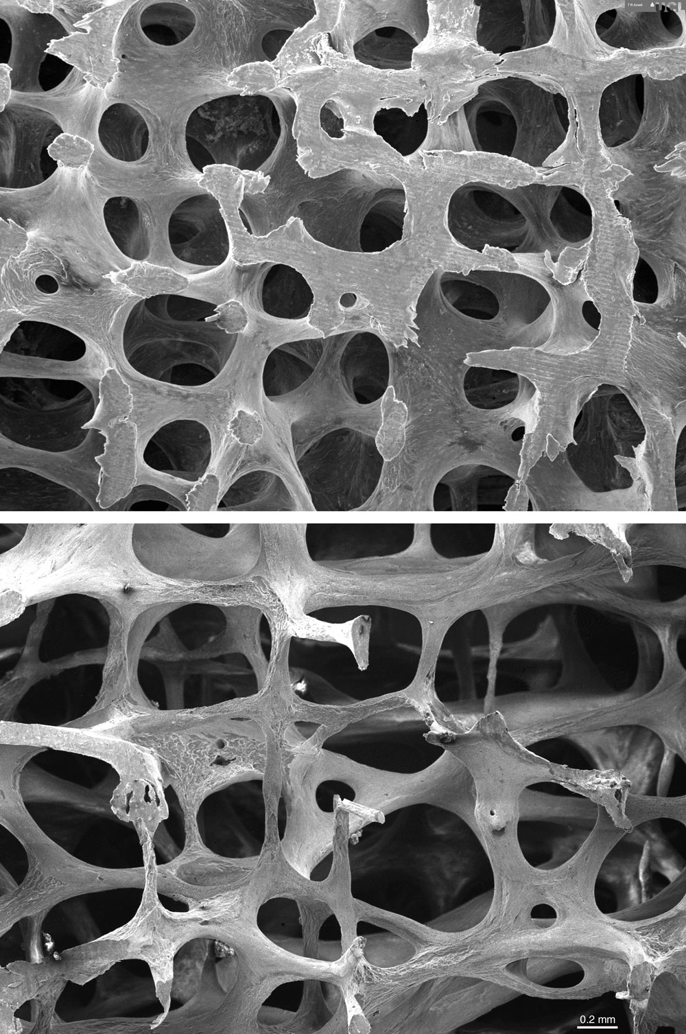
Osteoporotic Bone
Low power scanning electron microscope image, showing
osteoporotic architecture in the fourth lumbar vertebra
of an 89 year old woman (x20). The
bone is heavily eroded in places by the action of
osteoclasts and consists mainly of thin, fragile
struts. By kind permission of Alan Boyde

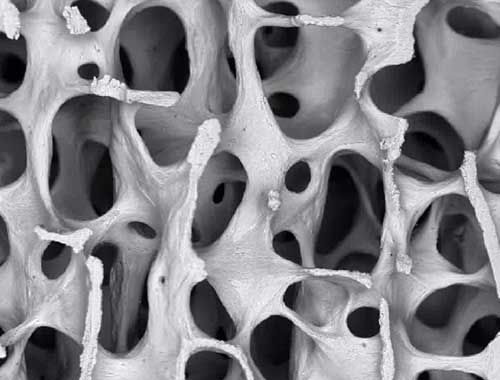
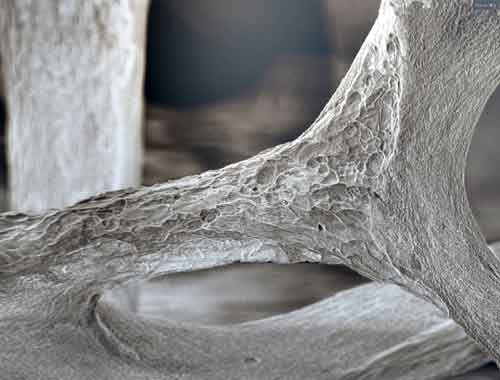
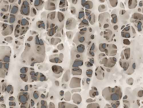
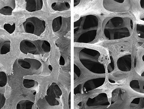
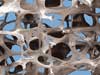
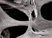
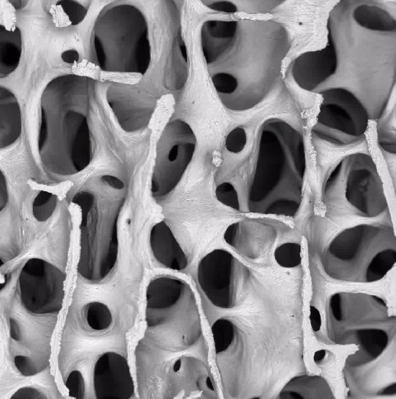
Normal Bone
Low power scanning electron microscope images,
showing normal bone architecture in the third lumbar
vertebra of a 30 year old woman (x20).
Strong, interconnected plates of bone are visible.
By kind permission of Alan Boyde
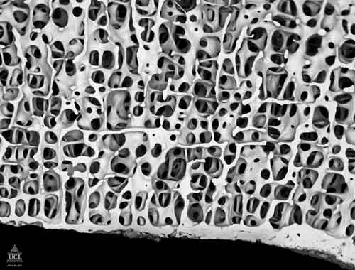
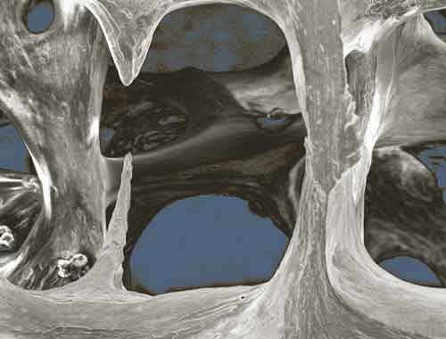
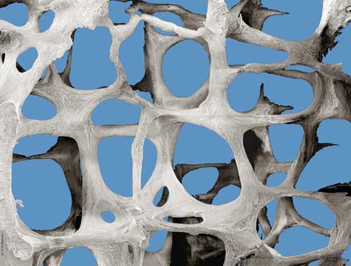
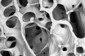
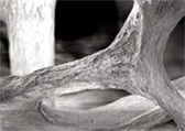
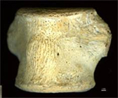
Normal Bone
Very low power scanning electron microscope image, showing
normal bone architecture in the fourth lumbar vertebra of
an 41 year year old man (x8). A regular pattern
of interconnected plates and thick struts of bone can be seen. By kind permission of Alan Boyde
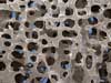
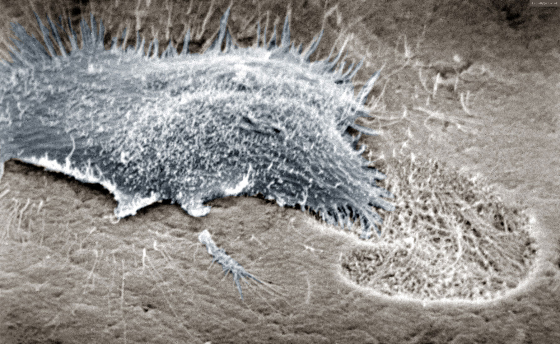
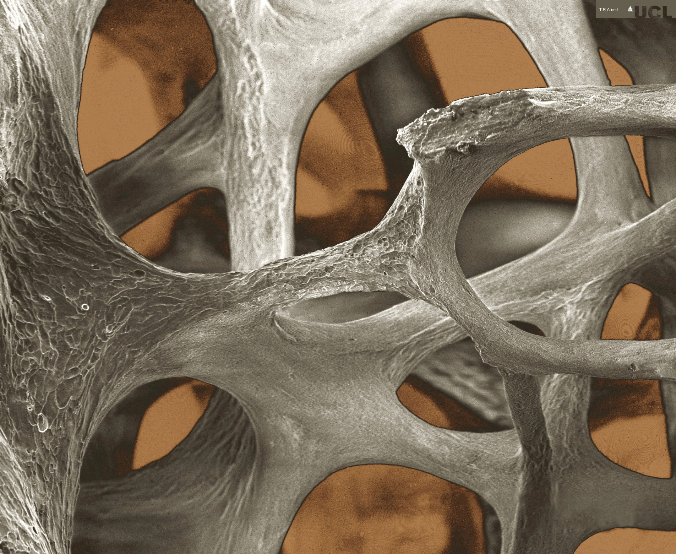
Osteoclast
Scanning electron micrograph showing osteoclast resorbing
bone. By kind permission of
Tim Arnett

Osteoclast
Breakfast, lunch and dinner for an osteoclast! By kind permission of Alan Boyde
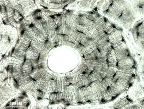
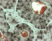
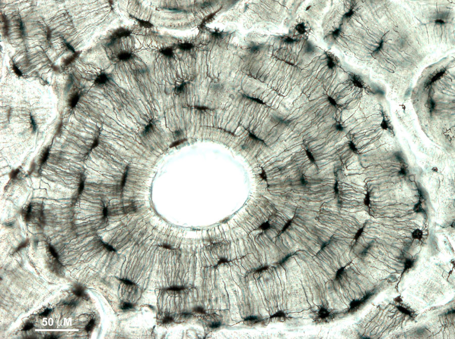
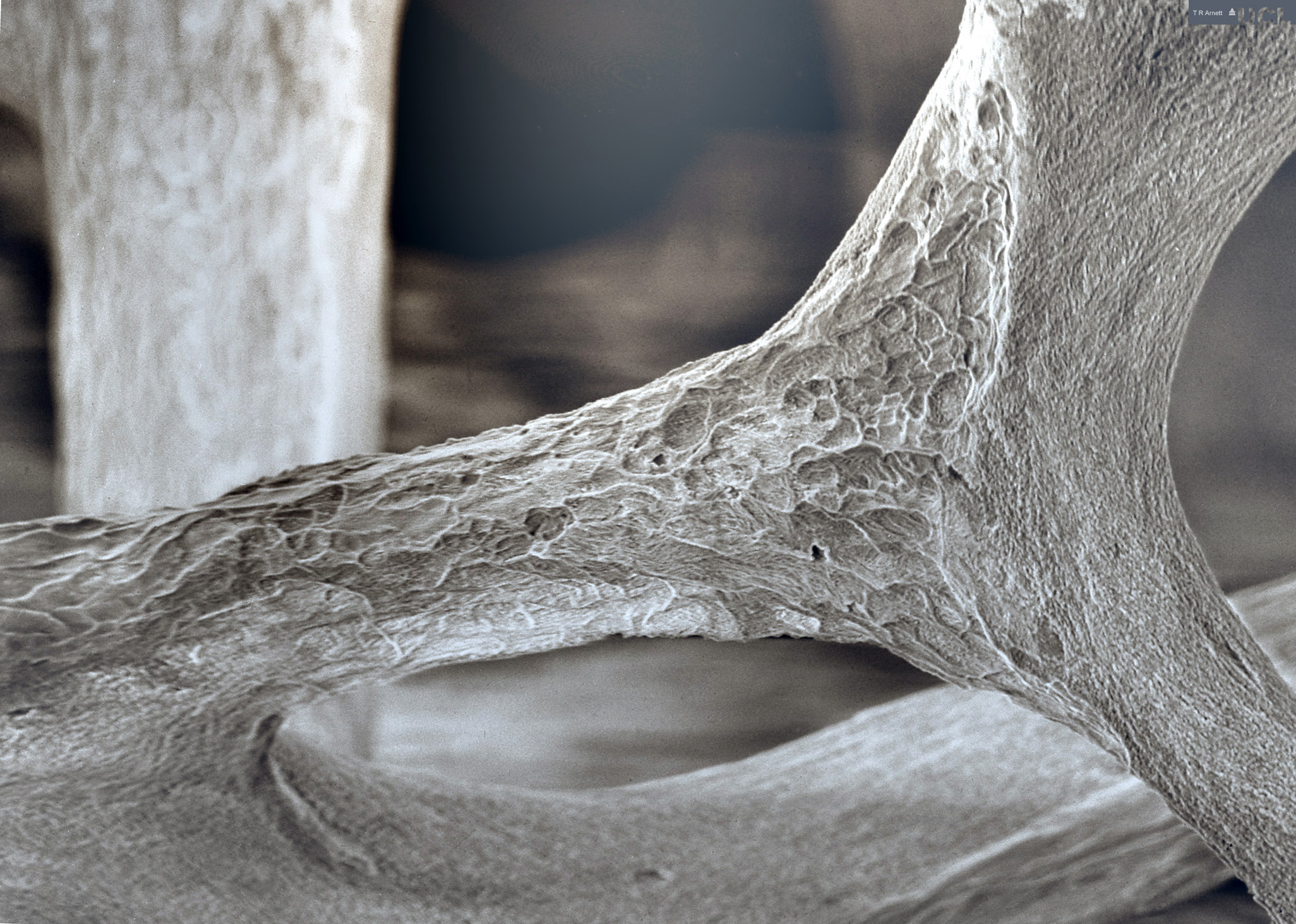
Osteocytes - Human Bone
Osteocytes - Human
bone. By kind permission of
Tim Arnett
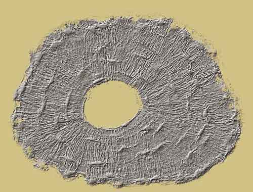
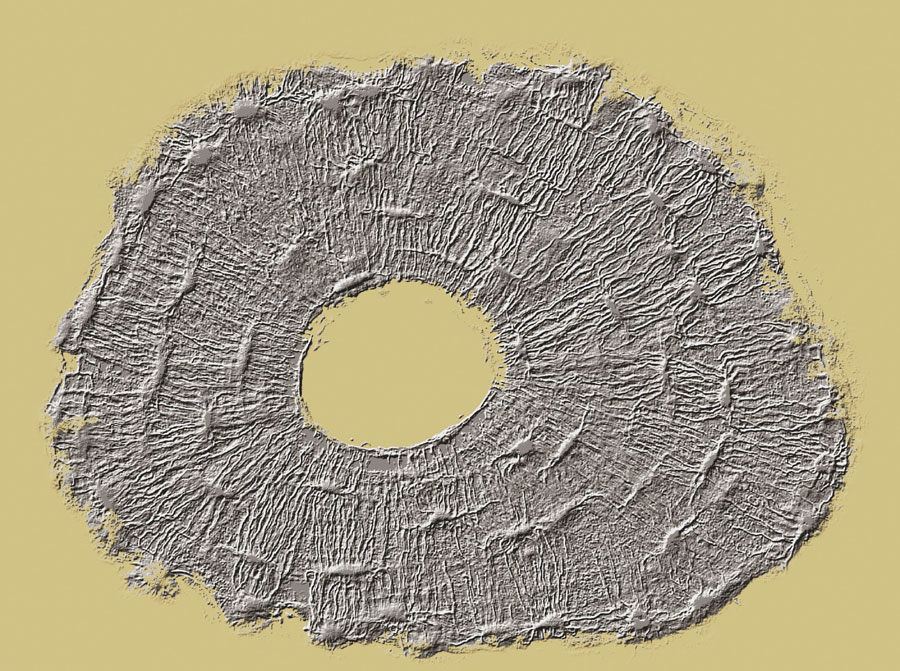
Osteocytes - Human Bone Osteon Relief
Osteocytes - Human
bone, osteon relief. By kind permission of
Tim Arnett
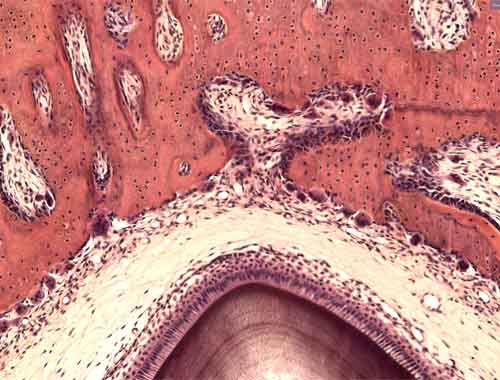
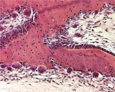
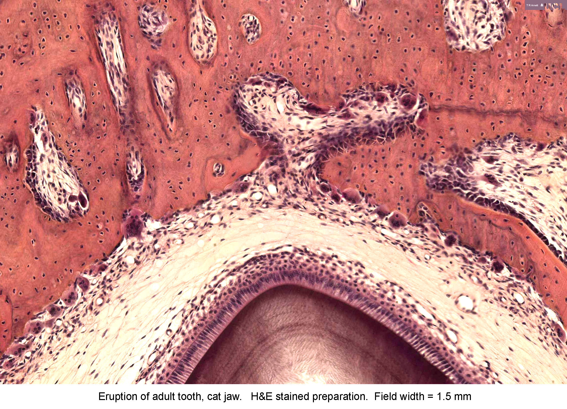
Tooth Eruption
Eruption of adult tooth, cat jaw. H & E stained preparation. Field view
1.5 mm. By kind permission of Tim Arnett
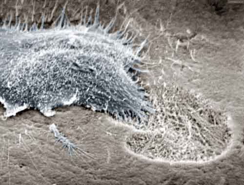
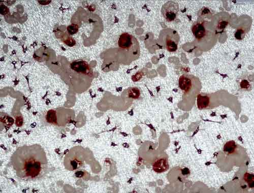
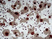
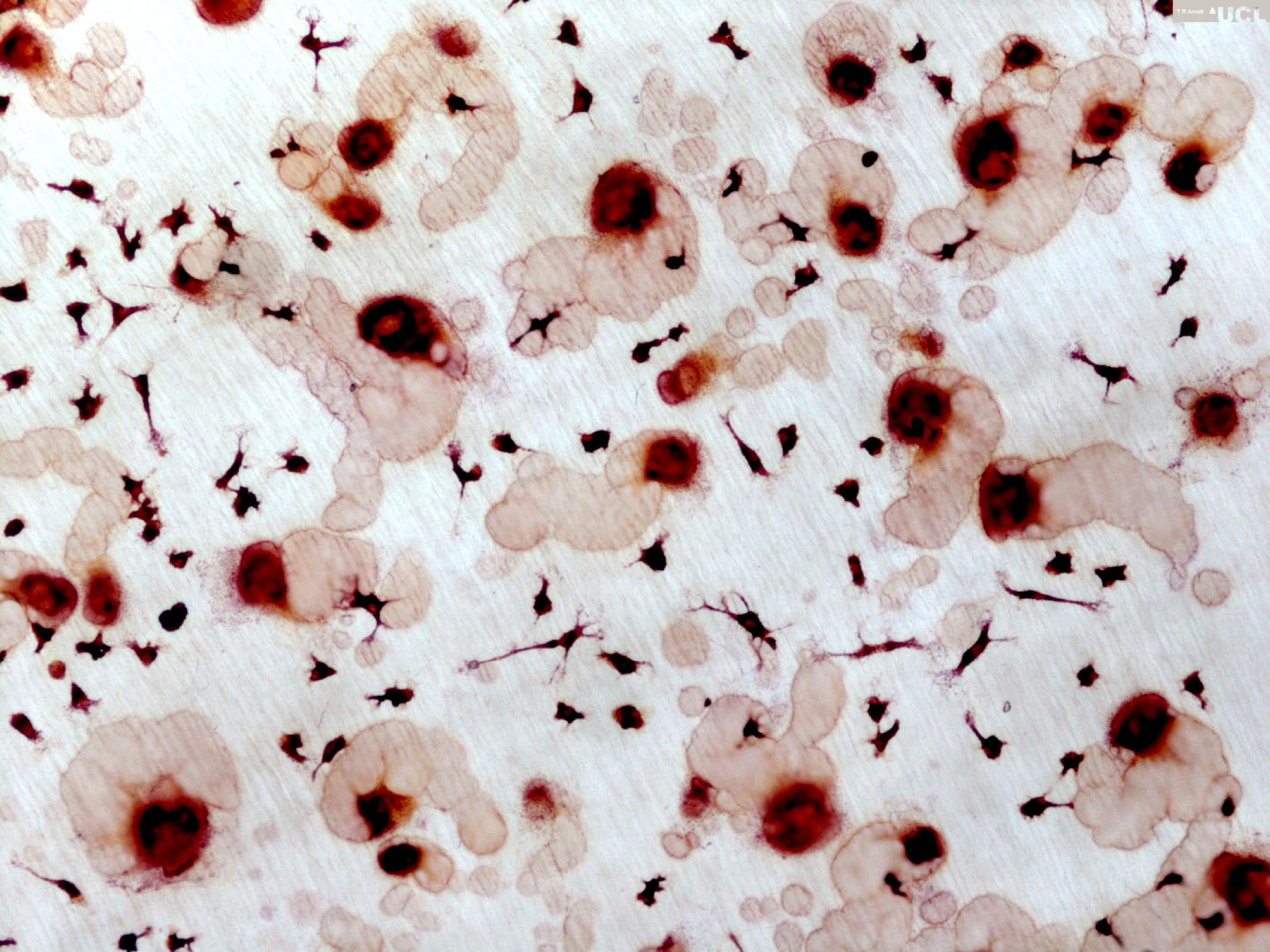
Human osteoclast & resorption trails #1
Human osteoclast & resorption trails #1. By
kind permission of Tim Arnett
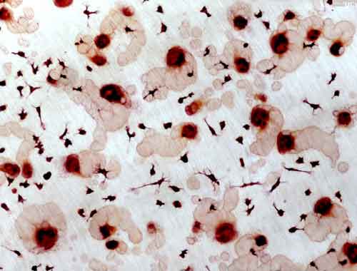
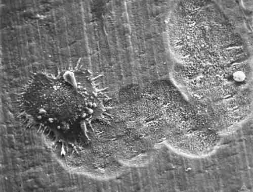
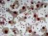
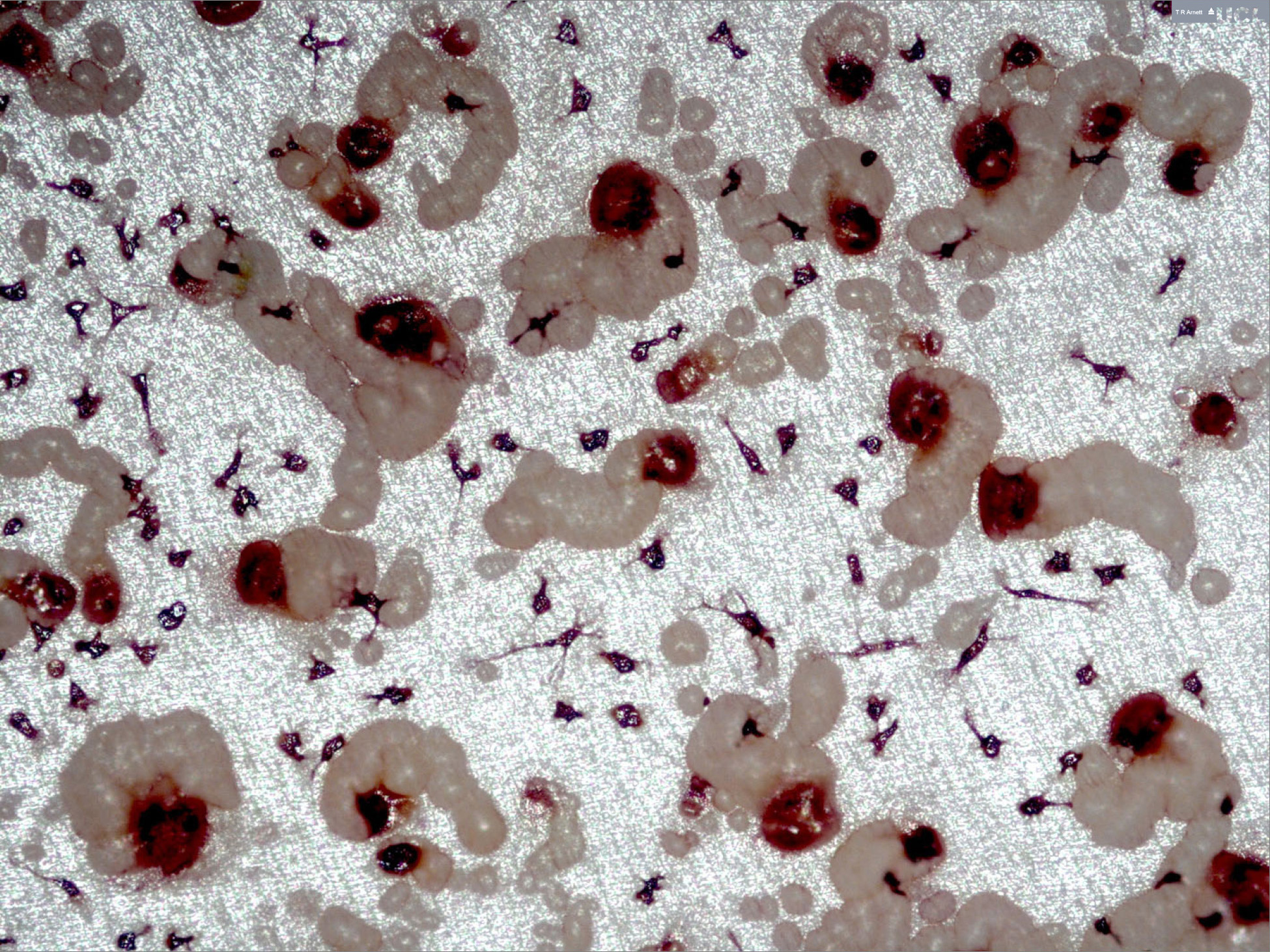
Human osteoclast & resorption trails #2
Human osteoclast & resorption trails #2. By
kind permission of Tim Arnett
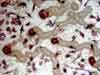
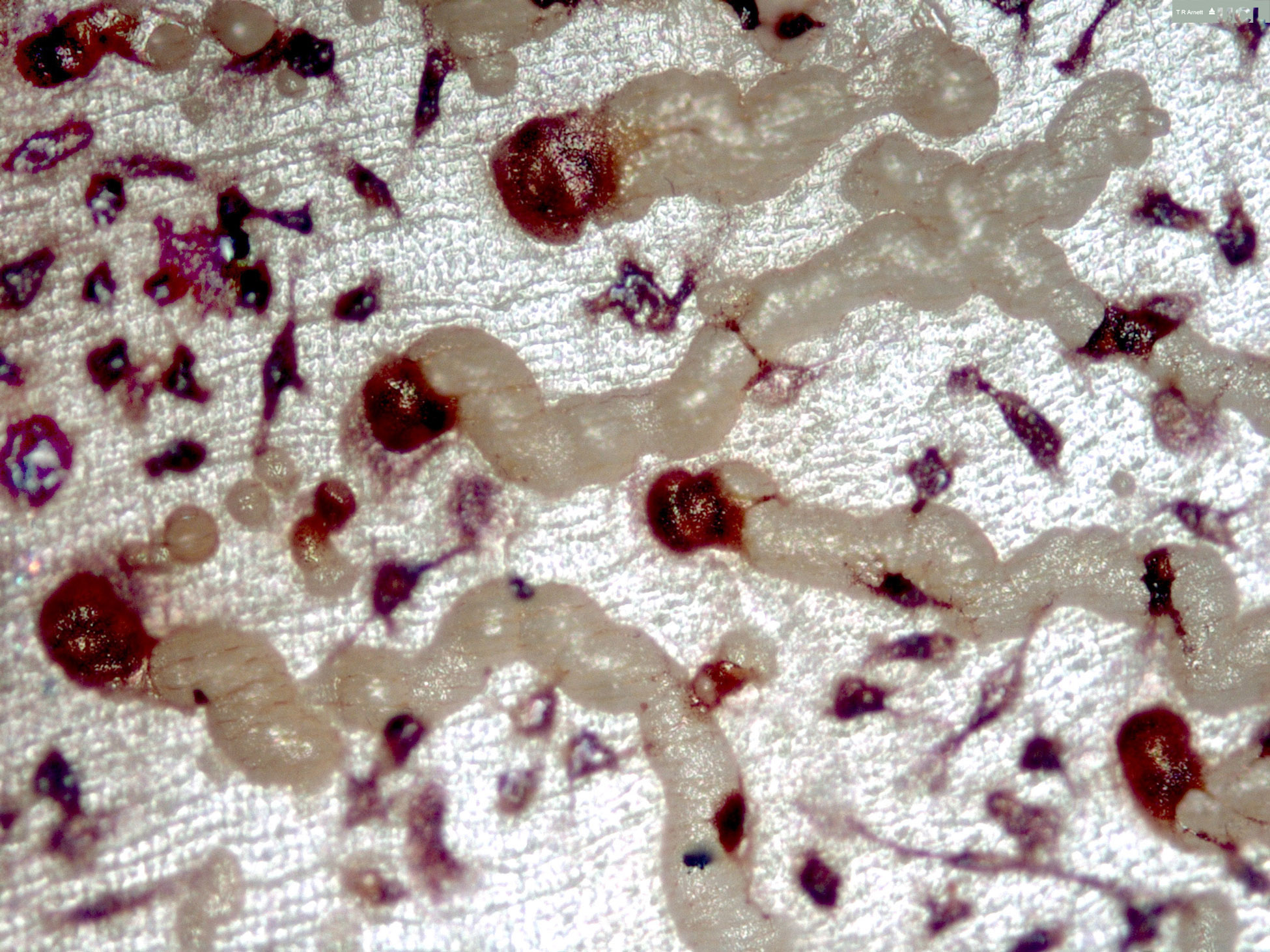
Human osteoclast & resorption trails #3
Human osteoclast & resorption trails #3. By
kind permission of Tim Arnett

Normal Bone
Image of normal bone architecture in the 3rd lumbar
vertebra of a 30 year old woman. By kind permission
of Tim Arnett.
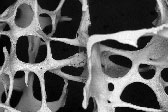
Osteoporotic Bone
Architecture in the 3rd lumbar vertebra of a 71
year old woman. Note trabecular bone element eroded
by osteoclasts. By kind permission of Tim Arnett.
Osteoporotic Bone
Architecture in the 3rd lumbar vertebra of a 71
year old woman. Note trabecular bone element perforated
by osteoclasts. By kind permission of Tim Arnett.
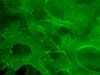
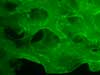
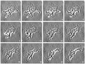
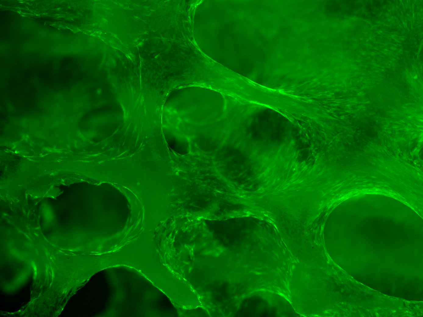
Human bone marrow stromal cells
Human bone marrow stromal cells spreading on trabecular bone. By kind permission
of Bram Sengers and Richard Oreffo

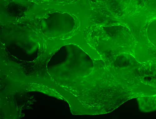
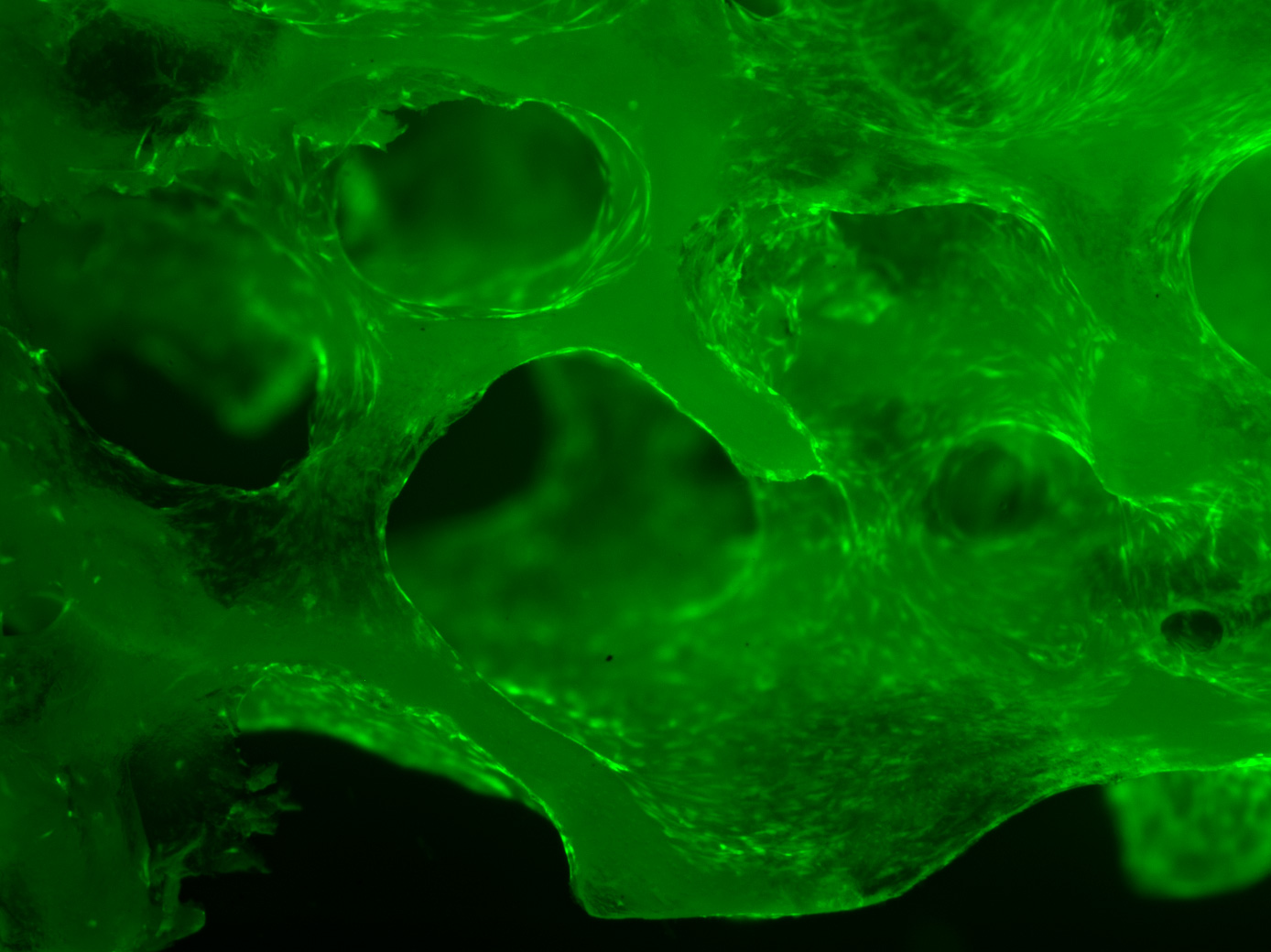
Human bone marrow stromal cells
Human bone marrow stromal cells spreading on trabecular bone. By kind permission
of Bram Sengers and Richard Oreffo
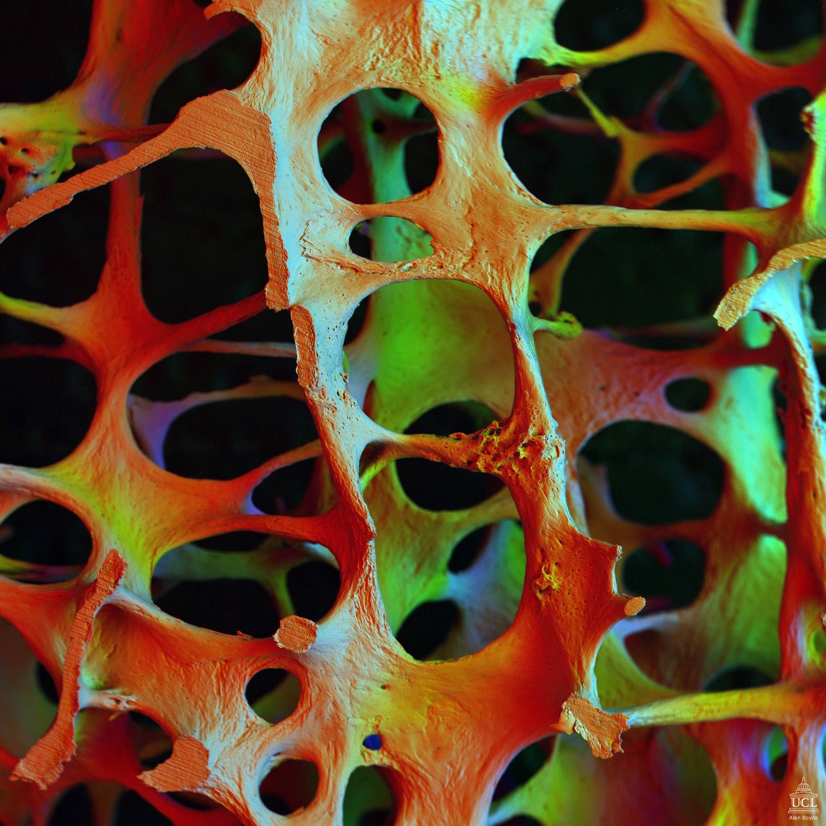
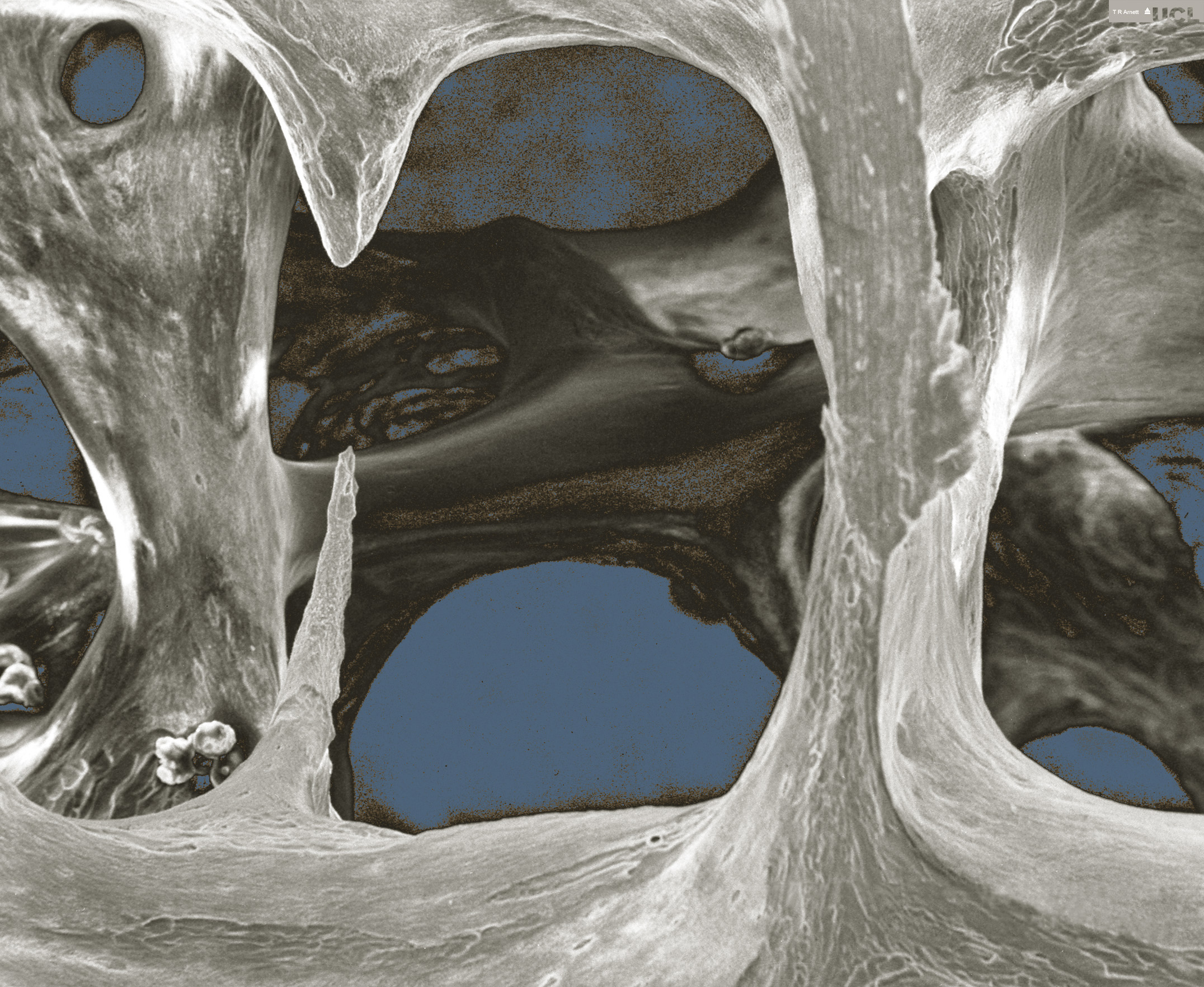
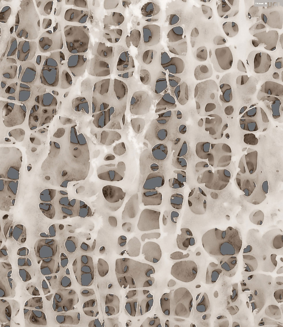
Osteoporotic bone architecture
Low-power scanning electron micrograph of osteoporotic
bone architecture in the 3rd lumbar vertebra of
a 71 yr old woman. Marrow and other cells have been
removed removed to reveal eroded bone elements.
Field width = 1.4 mm. By kind permission of Tim
Arnett.
Osteoporotic bone architecture
Low-power image of osteoporitic bone architecture.
By kind permission of Tim Arnett.
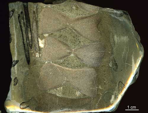
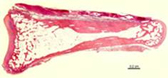
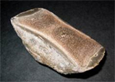
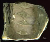
Section of ichthyosaur vertebrae and ribs
Section of ichthyosaur vertebrae and ribs in pyrite nodule. Seatown, Dorset, UK (190 myr). Trabecular bone structure is particularly well preserved in ribs. © Tim Arnett, University College London ([email protected]) High Resolution Version


