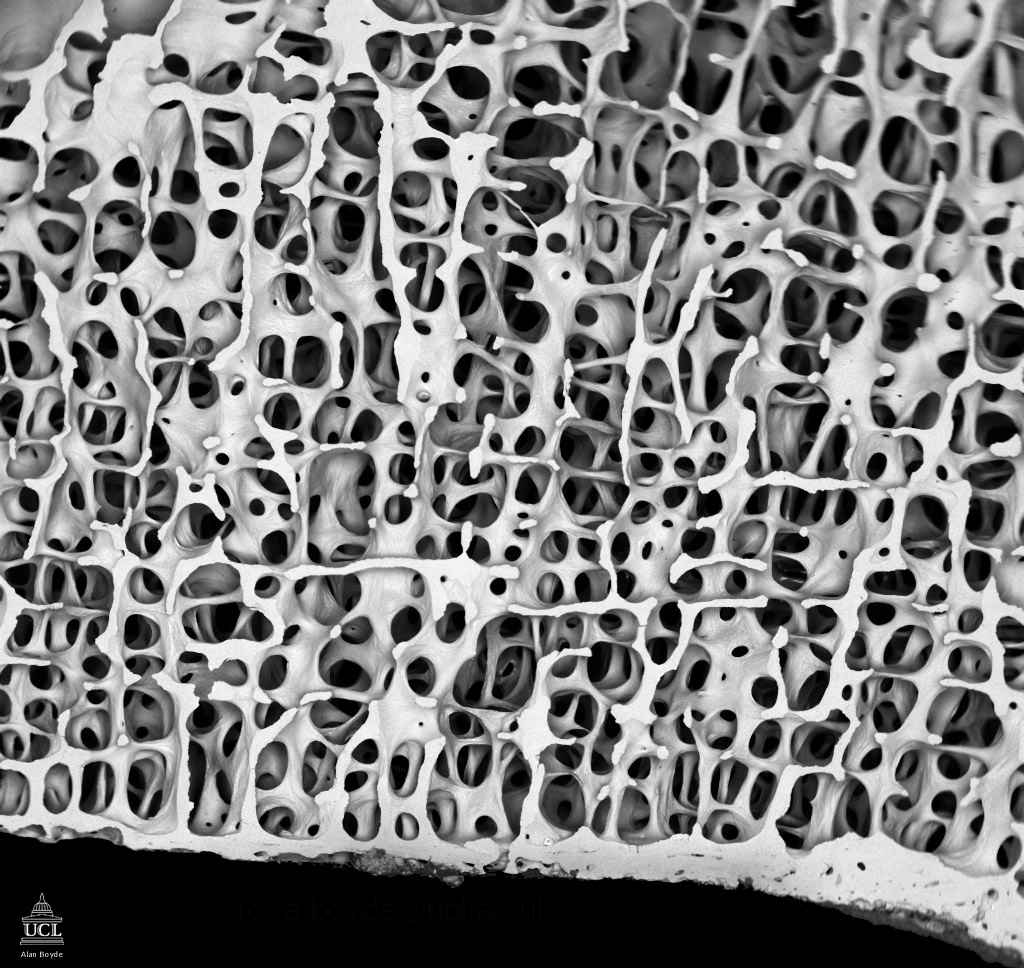
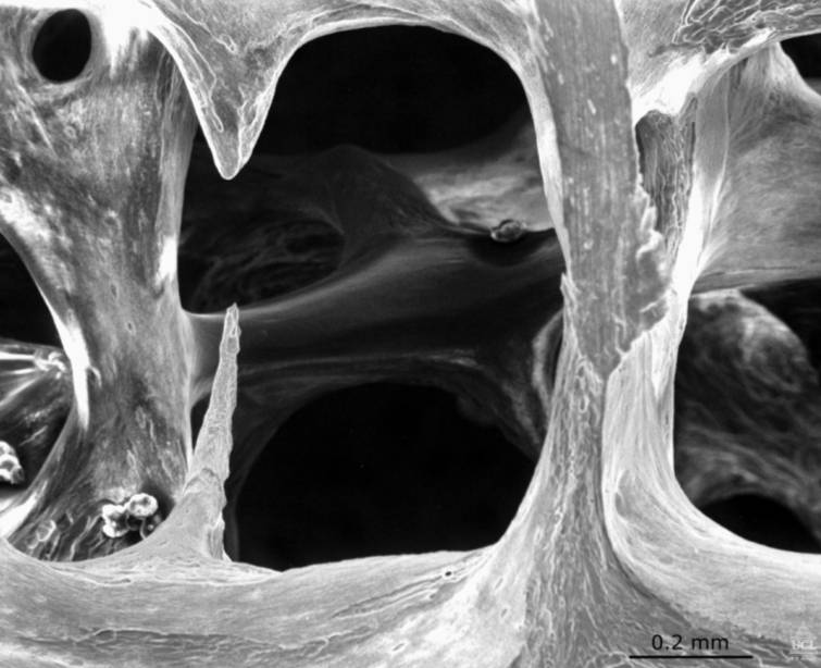
Osteoporotic bone
|
|
|
| |
enlarge image
|
|
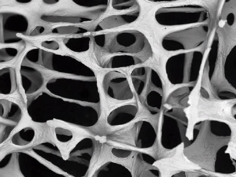
Low power scanning electron microscope
image, showing osteoporotic architecture in the fourth lumbar
vertebra of an 89 year old woman (20 times magnified). The bone
is heavily eroded in places by the action of osteoclasts and
consists mainly of thin, fragile struts. By
kind permission of
Alan Boyde ([email protected]) |
| |
|
|
|
| |
enlarge image |
|
View a video clip of osteoporotic bone
(of the vertebra described above)
Windows Media Player
(.wmv 0.6MB)
Quicktime Player (.mov
1MB)
By kind permission of
Alan Boyde ([email protected]) |
| |
|
|
|
Normal bone
|
|
|
| |
enlarge image
|
|
View a video clip of normal bone
Animation made from low power scanning electron microscope
images, showing normal bone architecture in the third lumbar
vertebra of a 30 year old woman (20 times magnified).
Strong, interconnected plates of bone are visible.
Windows Media Player
(.wmv 0.5MB)
Quicktime Player (.mov
0.5MB)
By kind permission of
Alan Boyde ([email protected]) |
| |
|
|
|
| |

enlarge image
|
|
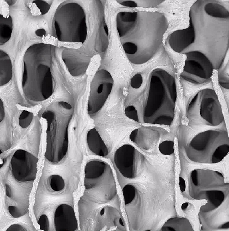
Very low power scanning electron microscope image, showing
normal bone architecture in the fourth lumbar vertebra of
an 41 year year old man (8 times magnified). A regular pattern
of interconnected plates and thick struts of bone can be seen.
By kind permission of
Alan Boyde ([email protected]) |
| |
|
|
|
Osteoclasts
|
|
|
| |

enlarge image
|
|
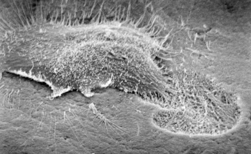
Scanning electron micrograph showing osteoclast resorbing
bone.
By kind permission of
Tim Arnett ([email protected]) |
| |
|
|
|
| |

enlarge image
|
|
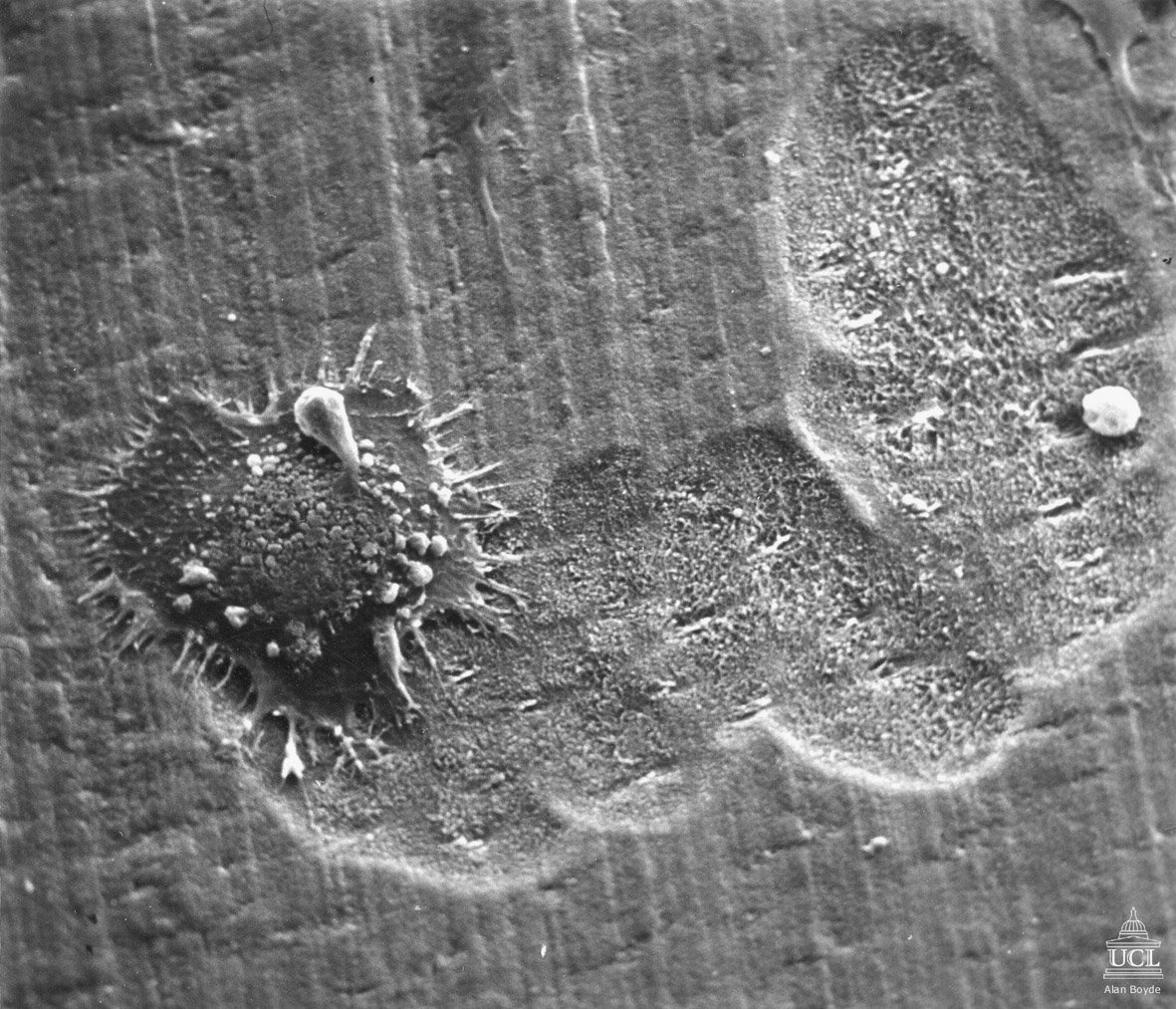
Breakfast, lunch and dinner for an osteoclast!
By kind permission of
Alan Boyde ([email protected]) |
| |
|
|
|
| |
Movie |
|
Osteoclast |
| |
 |
|
Time lapse movie of a rat osteoclast. This
large cell has 7 nuclei, and displays complex motile activity.
The sequence is speeded up 100 times
Windows Media Player
Low Resolution
(.wmv 3MB)
High Resolution
(.wmv 10MB)
Quicktime Player
Low Resolution
(.mov 5MB)
High Resolution
(.mov 9MB)
By kind permission of
Tim Arnett ([email protected]) |
| |
Movie |
|
Osteoblasts |
| |
 |
|
Time lapse movie of rat osteoblasts. This dense field
shows several hundred cells, including a few undergoing mitosis.
Note that many osteoblasts display large, waving processes,
resembling cilia. The sequence is speeded up 200 times.
Windows Media Player
Low Resolution
(.wmv 2MB)
High Resolution (.wmv
10MB)
Quicktime Player
Low Resolution
(.mov 4MB)
High Resolution (.mov
6MB)
By kind permission of Isabel Orriss ([email protected]).
|
| |
|
|
|
| Teaching slides |
|
|
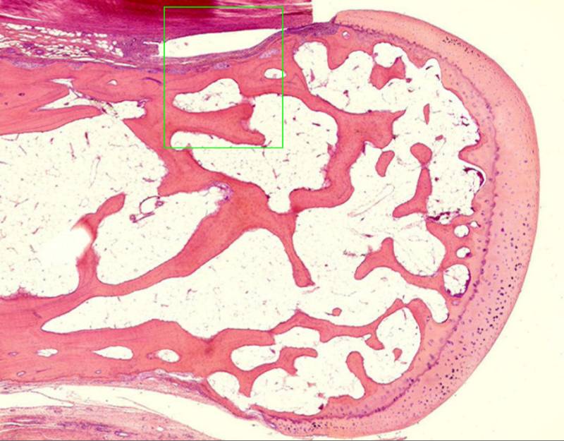
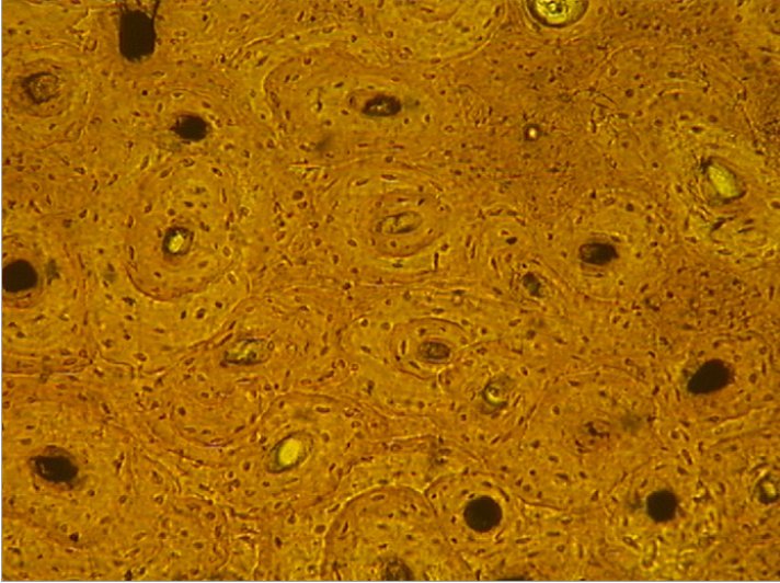
Bone Structure
|
|
|
| |

enlarge image
|
|
Animated sequence showing microscopic
structure of bone
(PPT slides 6.9MB)
PDF version 1.7MB
By kind permission of
Tim Arnett ([email protected]) |
| |
|
|
|
| |
enlarge image
|
|
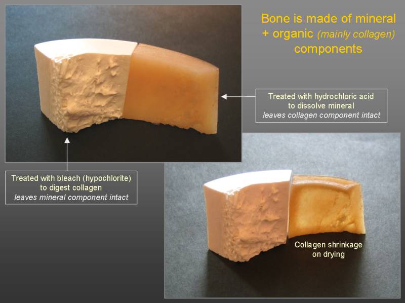
Composition of bone
(PPT slides 3MB)
PDF version 110K
By kind permission of
Tim Arnett ([email protected]) |
| |
|
|
|
| |
enlarge image |
|
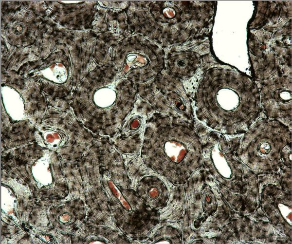
Cortical
bone animated sequences
(PPT slides 3MB)
PDF version
359K
By kind permission of
Tim Arnett ([email protected]) |
| |
|
|
|
| |
enlarge image |
|
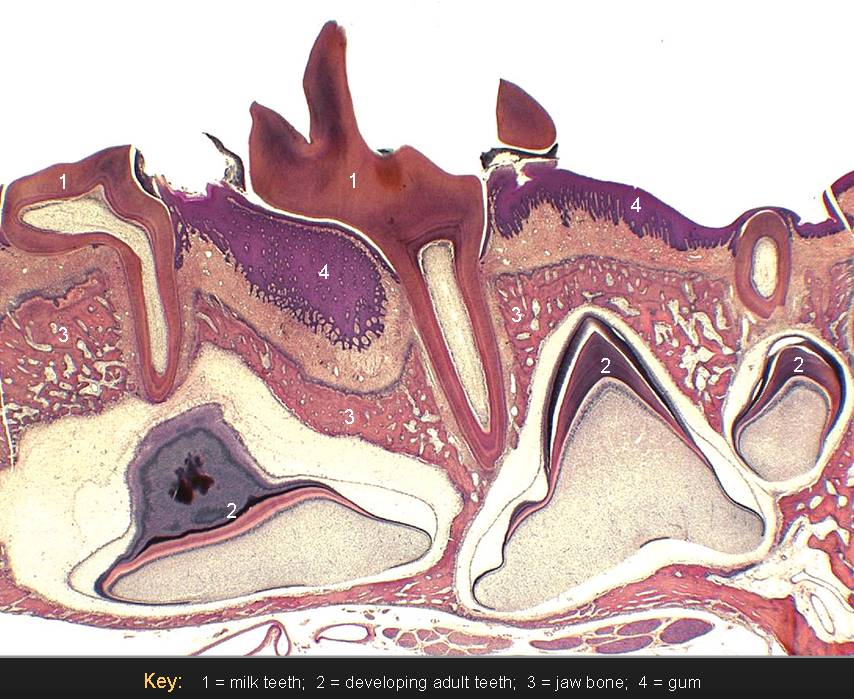
Tooth eruption
(PPT slides 3.3MB)
PDF version 982K
By kind permission of
Tim Arnett ([email protected]) |
| |
|
|
|
| |
enlarge image
|
|
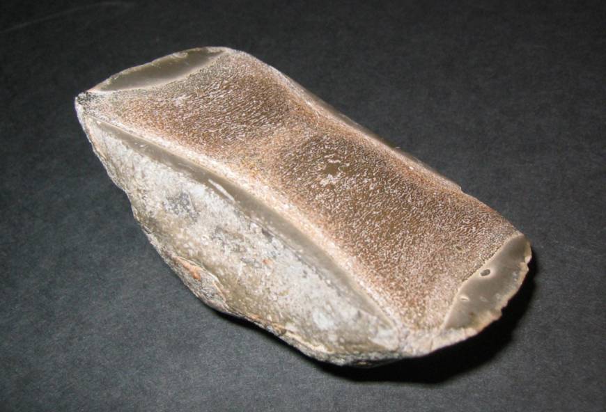
Ichthyosaur - fossil bone
(PPT slides 4.6MB)
PDF version 1.2MB
By kind permission of
Tim Arnett ([email protected]) |
| |
|
|
|
| |
 |
|
Iguanodon - fossil bone
(PDF 411K)
By kind permission of
Tim Skerry & John Currey
([email protected]) |
| |
enlarge
image |
|
|
| |
|
|
|
| |

enlarge image
|
|
Osteoporosis - bone erosion
by osteoclasts
(PPT slides 4MB)
PDF version 1.2MB
By kind permission of
Tim Arnett ([email protected])
|

